
Home / Journal / Oral Health and Dental Studies
Surgical Management of a Giant Compound Odontoma with 52 Denticles and Impacted Canine in the Maxilla of a Non-syndromic Child: A Case Report
Compound odontoma Denticles Supernumerary teeth Maxilla Non-syndromic Surgical management
Jayakumar Jayaraman
DOI: 10.31532/OralHealthDentStud.2.2.001 14 Jun 2021
Download

Abstract
Supernumerary tooth (ST) refers to any tooth or odontogenic structure that is formed excess of usual number for any given region of the dental arch. The treatment of ST usually involves surgical removal to avoid further complications to the developing dentition. This report presents a rare case of a giant compound odontoma that was observed in the left maxillary region of a 10-year-old child during routine dental examination. Surgical removal of the compound odontoma was performed in the dental setting under Nitrous oxide sedation. In total, 52 denticles were removed from the left maxillary region. Additionally, exposure, bonding, and orthodontic traction was performed in the contralateral impacted maxillary canine. Both surgical procedures were successful and well tolerated by the patient with adequate behavior management technique.
Keywords
Compound odontoma, Denticles, Supernumerary teeth, Maxilla, Non-syndromic, Surgical management
Introduction
Supernumerary tooth (ST) refers to any tooth or odontogenic structure that is formed excess of usual number for any given region of the dental arch. 1 The prevalence of ST varies between primary and permanent dentition, ranging from 0.3 to 0.6% in primary dentition to 3-6% in permanent dentition.2 Similarly, males have been reported to have a higher predilection compared to females. ST are mostly classified based on the location in the dental arch, morphology, and histological components.3 Around 90% of the ST has been reported in the anterior region of the maxilla, particularly in the palatal region. Amongst them, 80% are single tooth, with over 75% of the ST are impacted. 4 Based on the orientation, over 50% are normally oriented and 34% are inverted.4 Based on the size and composition, supernumerary teeth can be classified as complex, compound or mixed odontoma.5
The complications of ST include delayed or failure of eruption of permanent teeth, malocclusion, and ectopic eruption. When ST is in close proximity to permanent teeth, in some cases, loss of vitality and abnormal dental development have been reported.6 The diagnosis of ST is made during routine radiographic examination and mostly as an incidental finding. Majority of the ST remains innocuous in the primary dentition, and in some cases, throughout the life period.7 The severity of ST depends on its proximity to the normal set of dentition, anatomical structures, and complexity including the form and number.8 Genetic predisposition has been attributed as main reason for formation of ST with higher prevalence in Chinese ethnic populations.3 ST has been associated with several syndromes including Cleidocranial dysplasia, Gardner's syndrome, Ehler Danlos syndrome, Crouzon's syndrome, Down syndrome, and Ectodermal dysplasia.9,10 In non-syndromic patients, ST most commonly occurs as a single entity; however, presence of multiple ST has been reported in the literature.11,12
Several treatment options have been proposed for the management of ST that depends on interference of ST with the normal dentition, and presence or anticipated pathological conditions associated with the ST or adjacent teeth. For ST in the anterior region of the maxilla, 6 to 7 years has been established as an optimum age for surgical removal of the tooth.1 The authors found that beyond this age, the ST would cause damage to the permanent central incisors. In addition, it has been reported that dental development does not differ significantly between children with and without ST.13 Although ST could be diagnosed from panoramic radiograph or intra-oral periapical images using tube shift techniques, CBCT is usually recommended for surgical treatment planning.
Case Report
A 10-year-old Chinese male child presented to the outpatient clinic of School of Dentistry, International Medical University, Kuala Lumpur, Malaysia for routine dental examination. Medical history was non-contributory and hence established as American Society of Anesthesiologist (ASA) 1 category, not currently under any medications, and no known drug allergy. His Body Mass Index (BMI) was considered normal for his age and sex, and the vital signs were within normal limits. Dental history revealed presence of neonatal tooth #O, #P that were extracted immediately. The patient was previously seen by a general dentist who had performed amalgam restorations on primary molars.
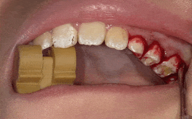
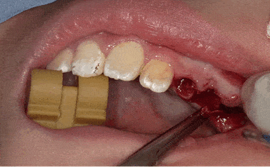
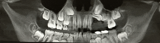
On clinical examination, the patient was in mixed dentition with primary canines and molars in the maxillary and mandibular arches. Universal numbering system was used to notate the teeth presented in this case report. Tooth #8 was hypoplastic with mild pitting (Figure 1). Amalgam restorations were found in tooth #S, #T. Patient had Class 1 molar and canine relationships with anterior deep bite. Panoramic radiographic was taken to assess his dental development stage, and incidentally, conglomerate mass of tooth structure was identified in the left side posterior maxillary region (Figure 2). A Cone Beam Computed Tomography (CBCT) was taken for better visualization that showed multiple ST above the roots of tooth #H, #I and #J with displacement of tooth #12, and #13 (Figure 3). Sagittal section of the CBCT showed displacement of tooth #11 and #12 (Figure 4). Based on the radiographic presentation, a diagnosis of compound odontoma in the left maxilla was made. Clinically, the lesion was asymptomatic, and the patient did not have pain during palpation in the left maxillary region. From the panoramic radiograph and CBCT, it was found that teeth #6 and #11 were impacted. The root of primary canines (tooth #C,H) were intact without any sign of resorption (Figure 2). Hence, in consultation with an Orthodontist, it was decided to perform a surgery for exposure and bonding of the tooth #6 through closed eruption technique. On the left side maxilla, tooth #11 was impacted and showed ectopic eruption due to presence of odontoma. Hence, a decision was made to surgically remove all the odontoma and allow spontaneous repositioning of tooth #11, 12, and 13. If required, surgery would be planned for orthodontic traction of impacted teeth at a later time. As a result, following treatment plan was devised: Restoration of dental caries in primary molars, surgical removal of ST along and evaluate the position of tooth #11, #12, and surgical exposure, bonding and orthodontic traction of tooth #6. Both the patient and the parent agreed to the proposed treatment plan and were highly motivated towards receiving the treatment.
Figure 1. Teeth in occlusion showing deep bite and hypoplastic tooth #8.
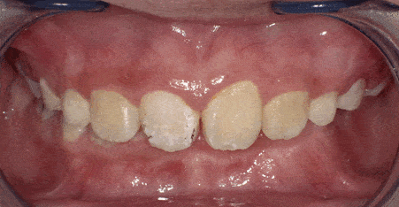
Figure 2. Panoramic radiograph taken during routine examination showing compound odontoma in the left side maxilla.
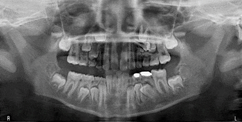
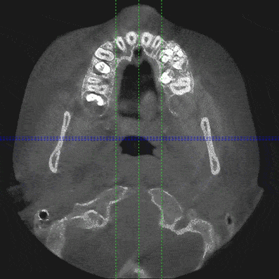
Figure 3. Axial slice CBCT scan showing compound odontoma in the left side maxilla.
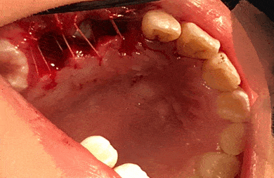
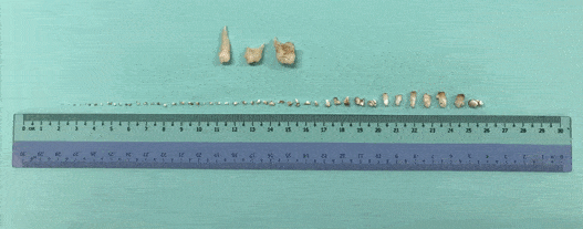
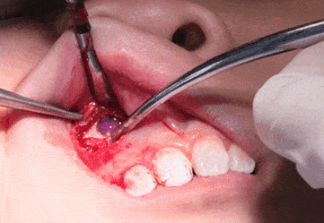
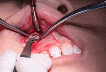
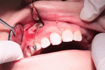
The patient was highly co-operative to dental treatment for restorations with Nitrous oxide sedation and his behavior was definitely positive (+/+) based on Frankl’s behavior rating scale. Considering his excellent behavior, it was planned to perform the surgery in the in-office dental setting with Nitrous oxide sedation alone. On the day of surgery, the patient was given Nitrous oxide and Oxygen at the ratio of 40:60 and his initial behavior was calm. Around 3.6 ml of 2% Lidocaine with 1:80000 Epinephrine was given as buccal and palatal infiltration in the left side maxillary posterior region. Tooth #H, I, J were extracted to allow access to the supernumerary teeth (Figure 5,6). After extraction of the primary teeth, ST were removed in sections. In addition to periosteal elevator, Warwick James elevator was used to remove the ST gently without damaging the adjacent permanent teeth. A follow up CBCT scan was taken immediately after the surgery to ensure that all ST were removed (Figure 7). The socket was then irrigated with normal saline, and 6 x 4.0 Polyglactin 910 (Vicryl, Ethicon) sutures were placed (Figure 8). Hemostasis was achieved and post-operative instructions were given including Acetaminophen 10mg/kg/dose in divided doses for pain Pro-Re-Nata (PRN), and soft diet for 2 days. An appointment was given after 2 weeks for recall evaluation. Post-operative phone call was made on the following day and the patient was found to be in stable condition. The extracted denticles were cleaned and numbered. In total, 52 denticles were extracted, and the size ranged from 0.2mm to 5mm in diameter. Some of the denticles were isolated but others were fused. Interestingly, six denticles had tuberculate appearance mimicking miniature incisors (Figure 9).
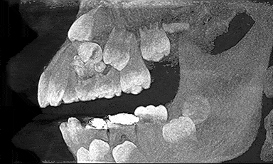
Figure 4. CBCT 3-D volumetric image in sagittal section showing compound odontoma and displacement of tooth #11 and #12.
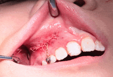
One month following the surgical removal of odontoma, the patient was scheduled for surgical exposure and bonding on tooth #6 for orthodontic traction. As with previous surgery, the Nitrous Oxide and Oxygen was placed at a ratio of 40:60. About 1.8 ml of 2% Lidocaine with 1:80000 Epinephrine was injected in the buccal mucosa of tooth #C and #7 region. A horizontal incision was made at the mucogingival junction of tooth #7 with full thickness mucoperiosteal exposure. The bone around the impacted tooth #6 was removed with No.8 surgical bur under copious saline irrigation. Tooth #6 was exposed and 0.5ml of 2% Lidocaine with 1:80000 Epinephrine was given for hemostasis (Figure 10). Once the bleeding was controlled, exposed tooth #6 was dried and etched with 37% orthophosphoric acid (Figure 11). The tooth was cleaned, and dried followed by application of prime and bond under strict isolation. A 3.5mm flat gold mesh button with chain was then bonded to tooth #6 with composite resin (Figure 12). The chain was then measured and attached to the orthodontic button on tooth #C using a ligature wire (Figure 13). Following verification of the attachments in tooth #6 and #C, flap was repositioned, and 7 x 4.0 Vicryl sutures were placed (Figure 14). Acetaminophen was prescribed for pain control and the patient was advised on a soft diet for 2 days. The patient was scheduled for recall appointment after 2 weeks for evaluation and initiation of comprehensive Orthodontic treatment.
Figure 10. Tooth #6 exposed following a full thickness mucoperiosteal incision near the mucogingival junction.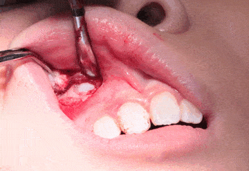
Discussion
To my understanding, this is the first ever case with fifty-two denticles extracted from the posterior maxilla. Although the overall prevalence of supernumerary teeth (ST) is estimated at 3-6%, single ST occurs in 76% to 86% of cases, and double ST occurs in 18% to 27% of cases.14 More than three ST is rare with an estimated prevalence of only 1% to 5%.15 Multiple ST is considered as a very rare phenomenon, particularly in non-syndromic patients and it tends to have a higher predilection to occur in the mandible compared to maxilla.12 The number of supernumerary teeth in non-syndromic patients are mostly reported in single numbers, however, as a rare entity, two studies reported 14 and 17 supernumerary teeth respectively.16,17 Similarly, only a few cases of compound odontoma with unusual size has been reported in non-syndromic patients. A literature review of odontoma based on sixty-one cases found that the number of odontoma ranged from 4 to 27.18 Sharma et al. reported a case of surgical removal of 37 denticles from anterior maxilla of a 17-year-old male child.19 Recently, Kunusoth et al. presented compound odontoma in a 20-year-old female with around 30 denticles and size ranging from 3mm to 9mm.20
As with most cases, this patient presented without symptoms and the compound odontoma was identified during routine radiographic examination. The decision to surgically remove the odontoma was made since it resulted in impaction of tooth #11 and deflection of tooth #12. The surgical approach to extract the odontoma was planned based on the CBCT images. Based on the location, extension and complexity, it was determined to remove them through the socket following extraction of tooth #H, I, and J. Complete removal of odontoma was verified with a CBCT scan taken immediately after the removal. In the absence of CBCT, this could be performed with intra-oral periapical radiographs with different angulations. Removal of odontoma in this patient would allow the possibility of spontaneous alignment, and eruption of the impacted tooth #11 and #12. Any delay in the surgical removal of the odontoma would have resulted to ectopic eruption of impacted teeth which would have further complicated the treatment outcome. To avoid space loss, trans-palatal arch space maintainer was planned between tooth #3 and #14.
Exposure and orthodontic traction are a treatment of choice of impacted tooth that has lost its potential for spontaneous eruption.21 Tooth #6 was not clinically palpable, and based on the CBCT, it was located above the root of tooth #7. Extraction of tooth #C would not result in normal physiological eruption, and hence exposure and bonding were planned. It was decided to extract tooth #C later based on the eruption of tooth #6. Since the premolars are unerupted, the gold button was temporarily bonded on the primary canine. The gold-plated chain was planned to be activated on the commencement of comprehensive Orthodontic treatment. Considering the complexity of the case, it might be argued that this patient would benefit from treatment with general anesthesia where all the treatment could be accomplished in one procedure. This option was presented to the parent but denied due to financial constraints. Moreover, the patient’s behavior was excellent during the examination, and dental restorations. Based on this initial assessment, it was determined to proceed the treatment with Nitrous oxide, and the outcome was successful for both surgeries.
Conclusions
This case report presents one of the rarest dental anomalies in a child where a giant compound odontoma with 52 denticles were diagnosed in the posterior maxilla as a part of routine dental examination. Surgical removal of all the denticles within the compound odontoma was presented along with exposure, bonding and orthodontic traction of contra-lateral impacted maxillary canine.
Acknowledgement
I would like to thank my alma mater, International Medical University, Kuala Lumpur, Malaysia where this surgery was performed. I also thank Dr. Ekta Priya and Dr. Sivakumar Arunachalam for their assistance in surgical and orthodontic treatment planning for this child.
Funding Information
This study did not receive any funding.
Conflict of Interest
The author declares no conflict of interest.
References
- Omer RS, Anthonappa RP, King NM. Determination of the optimum time for surgical removal of unerupted anterior supernumerary teeth . Pediatr Dent. 2010; 32: 14–20.
- Leco Berrocal MI, Martín Morales JF, Martínez González JM. An observational study of the frequency of supernumerary teeth in a population of 2000 patients. Med Oral Patol Oral Cir Bucal. 2007; 12: E134–8.
- Anthonappa RP, Omer RS, King NM. Characteristics of 283 supernumerary teeth in southern Chinese children. Oral Surg, Oral Med, Oral Pathol, Oral Radiol, Endod. 2008; 105: e48–54.
- Rajab LD, Hamdan MA. Supernumerary teeth: Review of the literature and a survey of 152 cases. Int J Paediatr Dent. 2002; 12: 244–54.
- Patchett CL, Crawford PJ, Cameron AC, Stephens CD. The management of supernumerary teeth in childhood – a retrospective study of practice in Bristol dental hospital, England and Westmead dental hospital, Sydney, Australia. Int J Paediatr Dent. 2001; 11:259–65.
- Parolia A, Kundabala M, Dahal M, Mohan M, Thomas MS. Management of supernumerary teeth. J Conserv Dent. 2011; 14: 221–4.
-
Akgun OM, Sabuncuoglu F, Altug A, Altun C. Non-syndrome patient with
bilateral supernumerary teeth: Case report and 9-year follow-up. Eur J Dent. 2013; 7: 123–6.
- Açikgöz A, Açikgöz G, Tunga U, Otan F. Characteristics and prevalence of non-syndrome multiple supernumerary teeth: A retrospective study. Dentomaxillofac Radiol. 2006; 35: 185–90.
- Lubinsky M, Kantaputra PN. Syndromes with supernumerary teeth. Am J Med Genet A. 2016; 170: 2611–6.
- Subasioglu A, Savas S, Kucukyilmaz E, Kesim S, Yagci A, Dundar M. Genetic background of supernumerary teeth. Eur J Dent. 2015; 9(1):153–158.
- Altunok Unlu N, Altan A, Altan H. Multidisciplinary Treatment of Mesiodens Causing Delayed Tooth Eruption and Follow-Up: Report of Two Cases. J Ped Dent. 2020; 6:46.
- Altan H, Akkoc S, Altan A. Radiographic characteristics of mesiodens in a non‐syndromic pediatric population in the Black Sea region. J Invest Clinical Dent. 2019; 10:e12377.
- Mallineni SK, Jayaraman J, Wong HM, King NM. Dental development in children with supernumerary teeth in the anterior region of maxilla. Clin Oral Invest. 2019; 23: 2987–94.
- Sood PB, Patil B, Godhi S, Shetty DC. Multiple supernumerary teeth and odontoma in the maxilla: A case report. Contemp Clin Dent. 2010; 1: 45–6.
- Santos TD, Silva ER, Faria AC, Mello Filho FV, Xavier SP. Multiple supernumerary teeth in a nonsyndromic 12-year-old female patient-A case report. Brazilian Dent J. 2014; 25:79–82.
- Diaz A, Orozco J, Fonseca M. Multiple hyperodontia: report of a case with 17 supernumerary teeth with non syndromic association. Med Oral Patol Oral Cir Bucal. 2009; 14:E229–E231.
- Altay MA, Ozgur B, Cehreli ZC. Management of a Compound Odontoma in the Primary Dentition. J Dent Child (Chic). 2016; 83: 98–101.
- Amado Cuesta S. Review of 61 cases of odontoma. Presentation of an erupted complex odontoma. Med Oral. 2003; 8: 366–373.
- Sharma U, Sharma R, Gulati A, Yadav R, Gauba K. Compound composite odontoma with unusual number of denticles – A rare entity. Saudi Dent J. 2010; 22: 145–149.
- Kunusoth R, Sahu V, Fathima A, Prakash R, S Pala SK, Maloth KN. Compound odontoma in the anterior maxilla. Oncol Radiotherapy. 2019; 46: 43–45.
- Chapokas AR, Almas K, Schincaglia GP. The impacted maxillary canine: a proposed classification for surgical exposure. Oral Surg Oral Med Oral Pathol Oral Radiol Endod. 2012; 113: 222–8.