Home / Journal / Oral Health and Dental Studies
Dento-Skeletal Abnormalities in School-aged Children in Iran: A Cross-Sectional Study
Preventive orthodontics dento-skeletal abnormalities malocclusion mixed dentition
Mohsen Shirazi, Yasamin Farajzadeh Jalali, and Hossein Hessari
DOI: 10.31532/OralHealthDentStud.2.1.003 12 Jan 2021
Abstract
Aim: The aim of this study was to evaluate skeleto-dental abnormalities in 9-11-year-old school children, in Tehran, Iran.
Materials and Methods: In this population-based cross-sectional descriptive study, a random cluster sampling was done among 19 school districts. A total of 1,429 socioeconomically and ethnically diverse Iranian schoolchildren, aged 9-11 years were studied. A brief questionnaire including background information such as gender and age was completed by the parents. Clinical examinations included the evaluation of sagittal and vertical skeletal relationship, Facial form (facial index), and the presence of significant asymmetry.
Results: There were 758 males and 671 females with the mean age of 10 years±8 months. According to the sagittal skeletal relationship, the most prevalent type was convex (63%) that presenting the skeletal Cl II jaw relation; followed by straight (32.9%); and then concave (4.1%). In the vertical skeletal relationship, 73.9% of the children had an average facial relationship, 18.4 % had a long face pattern; and 7.8% had a short face pattern. Regarding facial form in the frontal view, the most common was the average form (79.3%); followed by narrow (14%); and broad (6.7%). The prevalence of significant facial asymmetry was 15.2%.
Conclusion: The prevalence of dento-skeletal abnormalities were high. The majority of the Iranian schoolchildren, aged 9-11 years, had at least one dento-skeletal abnormality, even though it is commonly preventable.
Keywords
Preventive orthodontics, dento-skeletal abnormalities, malocclusion, mixed dentition
Introduction
A dento-skeletal abnormality is a common developmental abnormality that occurs due to the distortion of maxillary and/or mandibular development, which may greatly impact the positioning, alignment, and the health of the teeth1. These abnormalities have multifactorial etiologies including: genetic defects; nutritional deficiencies; behavioral habits leading to increased tension on the jaw; ethnicity; socioeconomic status; demographics and other environmental factors2,3. In addition, different studies have found a specific variation in the prevalence of skeleto-dental abnormalities in different ethnic populations4–8.
Dento-skeletal abnormalities can affect speech and mastication efficacy. Bruxism, dental trauma, and dental caries are significantly more prevalent in skeletal deformity cases compared to normal cases 9–12. Additionally, they may negatively impact on the health of young patients by leading to airway obstructions that contribute to sleep apnea, disturbing gastric pH, and affecting immune function.
Dento-skeletal abnormalities can adversely impact the psychological, emotional, and overall well-being and quality of life in children 13. Therefore, planning the orthodontic treatment requires understanding the patient’s baseline demographics and the prevalence of different skeleto-dental abnormalities among the population.
Thilander et al. evaluated orthodontic treatment needs in children 5–17 years of age and showed the 88% of children had some type of abnormality, and 20% of them greatly needed orthodontic treatments, in Colombia14. Keski-Nisula et al. estimated that the prevalence of malocclusion in Finland was 68%- 93% depending on the values of unacceptable parameters used15. Bittencourt showed the 85% of 6–10-year-old children had some sort of altered occlusion and 53% of them needed preventive orthodontic treatments16. Kaur et al. reported a high prevalence of malocclusion 88% among Indian adolescent aged 13–17 years17. Akbari et al. established a meta-analysis to evaluate the prevalence of malocclusion in Iranian children; they showed the prevalence of Class I, II, III malocclusion was 55%, 25%, and 6% respectively18. Eslamipour et al. found the prevalence of 5%, 52%, and 44% for Class I, II, and III sagittal skeletal pattern, respectively, among the patients who needed orthognathic surgery19. Eslamian et al. reported 68% and 32% Class III and II sagittal skeletal relationship, respectively among the patients who had orthognathic surgery 20.
If preventative dento-skeletal abnormalities are diagnosed, and treated early, their progression thus may be inhibited at an age early, leading to a decrease in the need of extensive challenging, costly treatments. The goal of this study was to evaluate dento-skeletal abnormalities by determining the sagittal relationship, vertical relationship, facial form, and significant asymmetry in school-aged children in Tehran, Iran.
Materials and Methods
Sample size calculation: The target population of the present study was Iranian children in mixed dentition before a growth spurt students in the fourth and fifth grades of elementary school in Tehran, Iran, between the ages of 9-11 years (10 years±8months) were selected as the study population.
To have a representative sample, cluster random sampling was applied. In each district, one boys’ school and one girls’ school were selected from a list of all schools in the district and in each school, students in grades four and five were selected. In 19 districts, a total of 38 schools were selected, and out of 1585 subjects, the data of 1429 children were collected (response rate = 90%).
Participation was voluntary and informed consent was obtained from the participants’ parents or legal guardians prior to conducting the study. The study was approved by the ethical committee of the school of dentistry, Tehran University of Medical Sciences (Ethical approval number: IR.TUMS.REC.1394.2003).
Study sample: The examiners were four orthodontists calibrated previously during the examination on 25 students by a professor of orthodontics (Kappa=0.95). Examinations were carried out in schools. A brief questionnaire including background information and a written anonymous consent form was given to the students to deliver to their parents to be completed and signed for participation in the study and further clinical examination. Clinical examinations included the most important skeletal indices described below.
Background information included gender, age (calculated by years and months and categorized as <120 months (<10 years), and >120 months (>10 years)).
Frontal view evaluation:
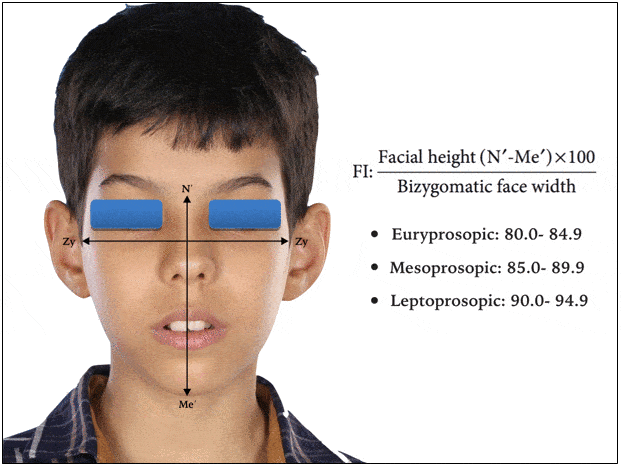
Facial Form: The facial form was recorded with facial index. It is an expression of the ratio between the facial height and the bi-zygomatic facial width. It is used in anthropometry to classify as Euryprosopic (Broad facial form) that is whom transversely wider, Mesoprosopic (Average facial form) that is the average and, Leptoprosopic (Narrow facial form) that is whom vertically relatively tall (figure 1)21.
Figure 1. Facial Index: facial height-to-width ratio

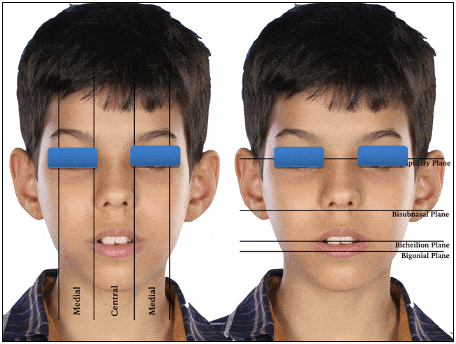
Significant asymmetry: Vertical asymmetry was recognized by the evaluation of parallelism of horizontal reference lines connecting the right and left pupillary, subnasale, corner of the mouth (Cheilion), and gonial angels. Horizontal asymmetry was recorded by the evaluation of proportionality of the widths of the face dividing into central, medial, and lateral equal fifths. The separation of the eyes and the widths of the eyes, which should be equal, determine the central and medial fifths (figure 2) 6.
Figure 2. Vertical and horizontal reference lines to evaluate asymmetry

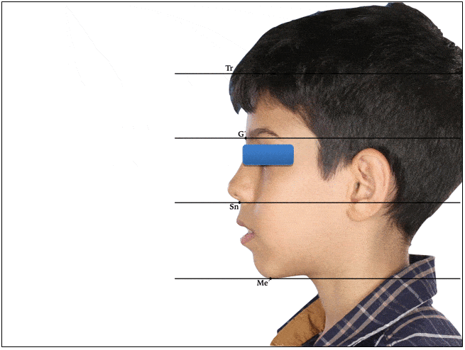
Vertical skeletal relationship: Vertical dimension was evaluated by examining the vertical facial thirds which measured the distance from the hairline to the base of the nose, base of the nose to the bottom of the nose, bottom of the nose to the chin. In an average face these measurements should be the same, If the lower third of the face is longer than the others, it is classified as an increased facial height (long face), and if it is shorter, it is classified as a decreased facial height (short face) (figure 3)5.
Figure 3. Horizontal reference lines to evaluate vertical height of the face

Profile view evaluation, sagittal skeletal relationship:
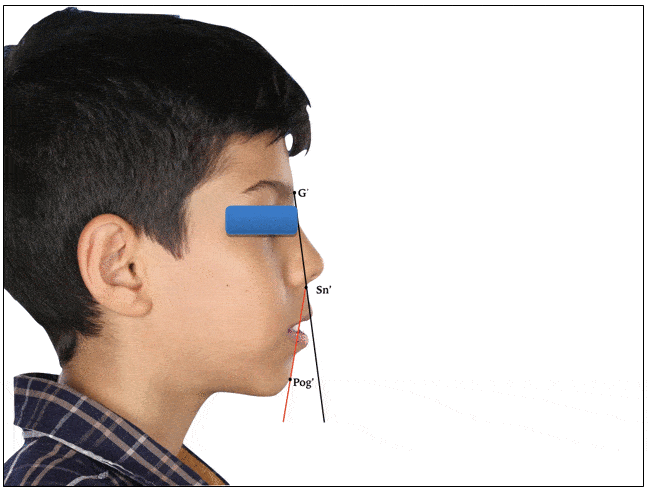
Analysis of profile type clinically by evaluating the soft tissue profile requires placing the patient in the natural head position. With the head in this position, the relationship between two lines are drawn, one dropped from the bridge of the nose to the base of the upper lip, and a second one extending from the point down to the chin; The relationship should ideally should form a nearly straight line, with only a slight inclination in either direction. A large angle between them indicates either profile convexity when upper jaw is prominent relative to the chin or profile concavity when the upper jaw is behind the chin. A straight profile indicates Class I jaw relationship, a convex profile indicates a skeletal Class II jaw relationship, and a concave profile indicates a skeletal Class III jaw relationship (figure 4)22.
Figure 4. Angle of facial profile convexity

Statistical analysis: Data were analyzed by SPSS version 20 (IBM, Chicago, USA). The Chi-square test was used to compare frequency between subgroups. P-values less than 0.05 were considered significant. Sagittal and vertical jaw relationship, facial form, and asymmetry were evaluated using binary logistic regression analysis.
Results
Out of a total of 1429 children, 758 (53%) were male and 671 (47%) were female.
The epidemiological information in percentage values on the prevalence of individuals with different facial form, discrepancies in sagittal and vertical plane and individuals with significant asymmetry was categorized.
According to the sagittal skeletal relationship, the most prevalent type was convex (63%) that present the skeletal Cl II jaw relationship, followed by straight (32.9%) and concave (4.1%). The straight profile is more prevalent (p<0.0001) in girls (38.8%) than boys (27.1%). The distribution of different types of sagittal skeletal relationship among different demographic features is given in Table 1.
Table 1. Weighted prevalence (%) of sagittal skeletal relationship among 9- to 11-year-old children (n=1429, Tehran, Iran, 2016)
|
Sagittal skeletal relationship |
||||
Straight (Cl I) |
Convex (Cl II) |
Concave (Cl III) |
P |
||
Gender |
Boys |
27.1 |
68.3 |
4.5 |
0.000* |
Girls |
38.8 |
57.7 | 3.6 |
||
Age(Year) |
9-10 |
32.4 |
63.7 |
3.9 |
0.770 |
10-11 |
33.4 |
62.2 |
4.4 |
||
|
Total |
32.9 |
63 |
4.1 |
|
*: P-value was less than 0.05. Results are weighted according to population of each district and boys/girl’s ratio.
In the vertical skeletal relationship, 73.9% of the children had a normal vertical relationship that was more prevalent (p<0.0001) in boys than the girls. 26.1% had some kind of altered relationship, so that, 18.4 % had a long face pattern and 7.8% had a short face pattern. The prevalence of normal face was decreased as the age increased. The distribution of different type of vertical skeletal relationship among different demographic features is given in Table 2.
Table 2. Weighted prevalence (%) of vertical skeletal relationship among 9- to 11-year-old children (n=1429, Tehran, Iran, 2016)
|
Vertical skeletal relationship |
||||
Normal |
Long face |
Short face |
P |
||
Gender |
Boys |
76.7 |
20 |
3.1 |
0.000* |
Girls |
70.8 |
16.7 | 12.5 |
||
Age(Year) |
9-10 |
75.4 |
16.9 |
7.6 |
0.298 |
10-11 |
72 |
20 |
8 |
||
|
Total |
73.9 |
18.4 |
7.8 |
|
*: P-value was less than 0.05.
Results are weighted according to population of each district and boys/girl’s ratio.
Regarding the facial form in frontal view, the most prevalent was average (79.3%) followed by narrow (14%) and broad (6.7%). The prevalence of average facial form in boys was lower (p=0.27) in boys (78.7%) than girls (79.9%). (Table 3).
Table 3. Weighted prevalence (%) of facial form among 9- to 11-year-old children (n=1429, Tehran, Iran, 2016)
|
Facial form |
||||
Average |
Broad face |
Narrow face |
P |
||
Gender |
Boys |
78.7 |
13 |
8.3 |
0.027* |
Girls |
79.9 |
14.9 | 5.1 |
||
Age(Year) |
9-10 |
78.4 |
14.4 |
7.2 |
0.575 |
10-11 |
80.3 |
13.5 |
6.1 |
||
|
Total |
79.3 |
14 |
6.7 |
|
*: P-value was less than 0.05.
Results are weighted according to population of each district and boys/girl’s ratio.
This study evaluated the significant asymmetry in vertical and horizontal direction but to summarize the vast amount of data we reported the asymmetry in general. Prevalence of asymmetry was 15.2%. As can be seen in Table 4 asymmetry was more prevalent (p=0.016) in boys (17.1%) than in girls (13.2%).
Table 4. Weighted prevalence (%) of symmetry among 9- to 11-year-old children (n=1429, Tehran, Iran, 2016)
|
Symmetry |
|||
Yes |
No |
P |
||
Gender |
Boys |
82.9 |
17.1 |
0.016* |
Girls |
86.8 |
13.2 | ||
Age(Year) |
9-10 |
85.3 |
14.7 |
0.329 |
10-11 |
84.4 |
15.6 |
||
|
Total |
84.8 |
15.2 |
|
*: P-value was less than 0.05.
Results are weighted according to population of each district and boys/girl’s ratio.
Discussion
Dento-skeletal abnormality is endemic and widespread throughout the world after dental caries and periodontal diseases and it is a significant public health burden on children. This demonstrates the great magnitude of the challenge that pediatric dentistry and orthodontics, in particular, need to confront.23 This study reports the prevalence of skeletal relationship in the sagittal, vertical, and transverse planes among 9-11-year-old school children.
In this study, we evaluated the sagittal skeletal relationship by a careful examination of the soft tissue of facial profile that yields the same information, though in less detailed for the underlying skeletal relationship. This was done with assessing the relationship between two lines, one dropped from the bridge of the nose to the base of the upper lip, and a second one extending from the point down to the chin that indicate the profile convexity and concavity. Profile convexity or concavity results from a disproportion in the size of the jaws so that a convex profile indicates a Class II jaw relationship and a concave profile indicates a Class III relationship6.
The above method has been introduced by Proffit WR and has been used previously to assess the sagittal skeletal relationship5,22.
The results showed that more than 60% of the 9-11 years old Iranian children had a convex profile, which was more prevalent in the boys than the girls. In contrast, the straight profile was more prevalent in girls than the boys. This may be related to the mandibular horizontal growth during the growth spurt and is more pronounced in girls during these years. Eslamipour et al, reported that the Class II skeletal relationship was the most prevalent (51.5%) among the patients who need orthognathic surgery19.
Different studies found similar variations in the occurrences of skeletal jaw relationships with respect to ethnicity. Jones found that the most common skeletal relationship among Saudi orthodontic patients was Class I followed by class II and Class III24. Al-Jundi and Riba showed the majority of Saudi Arabian patients were skeletal Class I followed by Class II and Class III25. Farawana found the Class I skeletal relationship as the most predominant in the Iraqi population, followed by Class II and Class III23. Zhou reported Class I jaw relationship as the most common relationships among the Chinese population followed by Class III and Class II26. Halwai showed the most common skeletal jaw relationship was skeletal Class II followed by Cl I and Cl III in Midwestern Nepal13.
Many studies described a variable prevalence of skeletal jaw relationships among Iranian children, all reporting the lowest level for skeletal Class III. The prevalence of dental malocclusion also reported by Borzabadi-Farahani and Akbari in Iranian children that they showed the most prevalent malocclusion was Cl I followed by Cl II, and the lowest was Cl III, although, the most prevalent malocclusion among the patients in need of orthognathic surgery was the Cl III malocclusion (45.6%)18,19,20,27. The variability among the different studies may be due to the varying methods and indices used to assess, differing sample selection, examiner subjectivity, and specific objectives.
This study showed that the 26.1% of children had some kind of altered vertical relationship, so that, the 18.4% of the children had a long face pattern and 7.8% had a short face pattern. Cardoso et al. reported the prevalence of 34.94% of vertical alteration and 14.06% of long face pattern in Brazilian children28. Woodside found 18% of long face in young Caucasian. In contrast29, Kelly reached 1.5% of long face pattern in American children30. This result showed the prevalence of facial form and asymmetry was significantly different in different gender, so that, the prevalence of broad facial form and asymmetry were more prevalent in boys than the girls.
The prevalence of average facial form in our study was 79.3%, narrow facial form was 14%, and the broad facial form was 6.7%. This study evaluated the significant asymmetry in vertical and horizontal direction. To summarize the high variant data, we reported the asymmetry in general. Of all children, 15.2% had degrees of asymmetry in their face. Anistoroaei et al concluded that facial asymmetry was present in 4.7% of patients, they also conducted that a significant correlation was evidenced between facial asymmetry and type of malocclusion, age, and type of dentition31.
However, Ferrario showed that no significant gender- or age-related differences were found for both metric and percentage indices of individual asymmetry32. Similar findings were reported by Farkas for adult males and females and by Burke for children aged 7–20 years33. Conversely, in a recent 3D study, the nose was more asymmetric in boys aged 9 years than in girls of corresponding age, although, mandibular asymmetry was greater in 6-year old boys than in girls of comparable age, no gender differences were observed in older individuals34. Studies conducted by Proffit and Sarver assessing facial asymmetries in orthodontic patients clinically found a prevalence of ranging 12-37% in the US, 23% in Belgium, and 21% in Hong Kong35. In Brazil, Boeck assessed the prevalence of the skeletal abnormalities. Their findings revealed a prevalence of 32% of asymmetry in their population36. Although both the methods of assessment and the definitions of malocclusions vary in different studies, and their findings should be compared with caution, the present results do not seem to significantly differ from those reported earlier.
The results obtained in this study revealed that the prevalence of dento-skeletal abnormalities was high and the majority of the Iranian school aged children, 9–11 years, have at least one dento-skeletal abnormality, even though it is commonly preventable.
References
- Mosca G, Grippaudo C, Marchionni P, Deli R. Class III dento-skeletal anomalies: rotational growth and treatment timing. Eur J Paediatr Dent: official journal of European Academy of Paediatric Dentistry. 2006 Jan;7(1):23–38.
- Aliaga-Del Castillo A, Vilanova L, Miranda F, Arriola-Guillén LE, Garib D, Janson G. Dentoskeletal changes in open bite treatment using spurs and posterior build-ups: A randomized clinical trial. Am J Orthod Dentofacial Orthop. 2020 Nov 18.
- Fabozzi FF, Nucci L, Correra A, d'Apuzzo F, Franchi L, Perillo L. Comparison of two protocols for early treatment of dentoskeletal Class III malocclusion: Modified SEC III versus RME/FM. Orthodontics & Craniofacial Research. 2020 Nov 11.
- Garner LD, Butt MH. Malocclusion in black Americans and Nyeri Kenyans. Malocclusion in black Americans and Nyeri Kenyans. Angle Orthod. 1985 Apr;55(2):139–146.
- Proffit WR, White R, Sarver DM, Contemporary treatment of dentofacial deformity. St. Louis, Mosby; 2003. pp. 158–167.
- Mouakeh M. Cephalometric evaluation of craniofacial pattern of Syrian children with Class III malocclusion. Am J Orthod Dentofacial Orthop. 2001 Jun;119(6):640–649.
- Siriwat PP, Jarabak JR. Malocclusion and Facial Morphology: Is There a Relationship? An Epidemiologic Study. Angle Orthod. 1985 Apr;55(2):127–138.
- Willems G, De Bruyne I, Verdonck A, Fieuws S, Carels C. Prevalence of Dentofacial Characteristics in Belgian Orthodontic Population. Clin Oral Investig. 2001 Dec;5(4):220–226.
- Martins-Junior PA, Marques LS, Ramos-Jorge ML. Malocclusion: social, functional and emotional influence on children. J Clin Pediatr Dent. 2012 Sep;37(1):103–108.
- Baskaradoss JK, Geevarghese A, Roger C, Thaliath A. Prevalence of malocclusion and its relationship with caries among school children aged 11 - 15 years in southern India. Korean J Orthod. 2013 Feb;43(1):35–41.
- Ghafournia M, Hajenourozali Tehrani M. Relationship between Bruxism and Malocclusion among Preschool Children in Isfahan. J Dent Res Dent Clin Dent Prospects. 2012 Dec;6(4):138–142.
- Bendgude V, Akkareddy B, Panse A, Singh R, Metha D, Jawale B, Garcha V. Et al. Correlation between dental traumatic injuries and overjet among 11 to 17 years’ Indian girls with Angle's class I molar relation. J Contemp Dent Pract. 2012 Mar;13(2):142–146.
- Halwai HK, Gautam V, Pandey M. Distribution of Different Skeletal Pattern in Patients Seeking Orthodontic Treatment in Mid-Western. Orthod J Nepal. 2017 May;6(2):7-9.
- Thilander B, Pena L, Infante C, Parada SS, de Mayorga C. Prevalence of malocclusion and orthodontic treatment need in children and adolescents in Bogota, Colombia. An epidemiological study related to different stages of dental development. Eur J Orthod. 2001 Apr 1;23(2):153-68.
- Keski-Nisula K, Lehto R, Lusa V, Keski-Nisula L, Varrela J. Occurrence of malocclusion and need of orthodontic treatment in early mixed dentition. Am J Orthod Dentofacial Orthop. 2003 Dec 1;124(6):631–638.
- Bittencourt MA, Machado AW, An Overview of the Prevalence of Malocclusion In 6 To 10-Year Old Children in Brazil. Dental Press J Orthod. 2010 Dec; 15(6): 113-122.
- Kaur H, Pavithra US, Abraham R. Prevalence of malocclusion among adolescents in South Indian population. J Int Soc Prev Community Dent. 2013 Jul;3(2):97.
- Akbari M, Lankarani KB, Honarvar B, Tabrizi R, Mirhadi H, Moosazadeh M. Prevalence of malocclusion among Iranian children: A systematic review and meta-analysis. Dent Res J. 2016 Sep;13(5):387.
- Eslamipour F, Borzabadi-Farahani A, Le BT, Shahmoradi M. A retrospective analysis of dentofacial deformities and orthognathic surgeries. Ann Maxillofac Surg. 2017 Jan;7(1):73.
- Eslamian L, Borzabadi-Farahani A, Badiee MR, Le BT. An Objective Assessment of Orthognathic Surgery Patients. J Craniofac Surg. 2019 Nov 1;30(8):2479-82.
- Naini FB, Gill DS, Orthognathic Surgery: Principle, Planning and. Chichester, West Sussex: Wiley-Blackwell; 2011. P. 877.
- Borzabadi‐Farahani A, Borzabadi‐Farahani A, Eslamipour F. An investigation into the association between facial profile and maxillary incisor trauma, a clinical non‐radiographic study. Dent Traumatol. 2010 Oct;26(5):403-8.
- Farawana NW. Malocclusion in Iraq. Quintessence Int. 1987 Feb; 18(2):153–157.
-
Jones Wb. Malocclusion and Facial Types in A Group of Saudi Arabia
Patients Referred for Orthodontic Treatment: A Preliminary Study. Br J Orthod 1987 Jul; 14(3):143–146.
- Al-Jundi A, Riba H. Pattern of Malocclusion in A Sample of Orthodontic Patients from A Hospital in The Kingdom of Saudi Arabia. Savant J Med & Medical Sci. 2015 Jun; 1(1):14–21.
- Zhou L, Mok Cw; Hagg U, Mcgrath C, Bendeus M, Wu J. Anteroposterior Dental Arch and Jaw-Base Relationships in A Population Sample. Angle Orthod. 2008 Nov; 78(6):1023–1029.
- Borzabadi-Farahani A, Borzabadi-Farahani A, Eslamipour F. Malocclusion and occlusal traits in an urban Iranian population. An epidemiological study of 11-to 14-year-old children. Eur J Orthod. 2009 Oct 1;31(5):477–484.
- Cardoso MD, Capelozza Filho L, An TL, Lauris JR. Epidemiology of long face pattern in schoolchildren attending fundamental schools at the city of Bauru-SP. Dent Press J Orthod. 2011 Apr;16(2):108–119.
- Woodside DG, Linder-Aronson S. The channelization of upper and lower anterior face heights compared to population standard in males between ages 6 to 20 years. Eur J Orthod. 1979 Jan 1;1(1):25–40.
- Kelly JE, Harvey CR. An assessment of the occlusion of the teeth of youths 12-17 years, United States. 1977.
- Anistoroaei D, Golovcencu L, Săveanu IC, Zegan G, The Prevalence of Facial Asymmetry in Preorthodontic Treatment. Int J Med Dent. 2014 Sep; 4(3):210–215.
- Ferrario VF, Sforza C, Miani A, Tartaglia G. Craniofacial morphometry by photographic evaluations. Am J Orthod Dentofacial Orthop. 1993 Apr; 103:327-337.
- Farkas LG, Cheung G, Facial asymmetry in healthy North American Caucasians. Angle Orthod. 1981 Jan; 52:70–77.
- Melnik, Andrew K, A cephalometric study of mandibular asymmetry in a longitudinally followed sample of growing children, Am J Orthod Dentofacial Orthop. 1992 Apr;101(4): 355 – 366.
- Thiesen G, Gribel BF, Freitas MP. Facial asymmetry: a current review. Dental Press J Orthod. 2015 Nov;20(6):110–125.
- Boeck Em, Lunardi N, Pinto As, Pizzol Kec, Boeck Neto Rj. Occurrence of Skeletal Malocclusions in Brazilian Patients with Dentofacial Deformities. Braz Dent J. 2011;22(4):340–345.


